Victory Laboratory & Ultrasound Services provides a range of ultrasound examinations to support the assessment and monitoring of different medical conditions. Our ultrasound services are designed to provide clear diagnostic information to assist healthcare professionals in patient care.
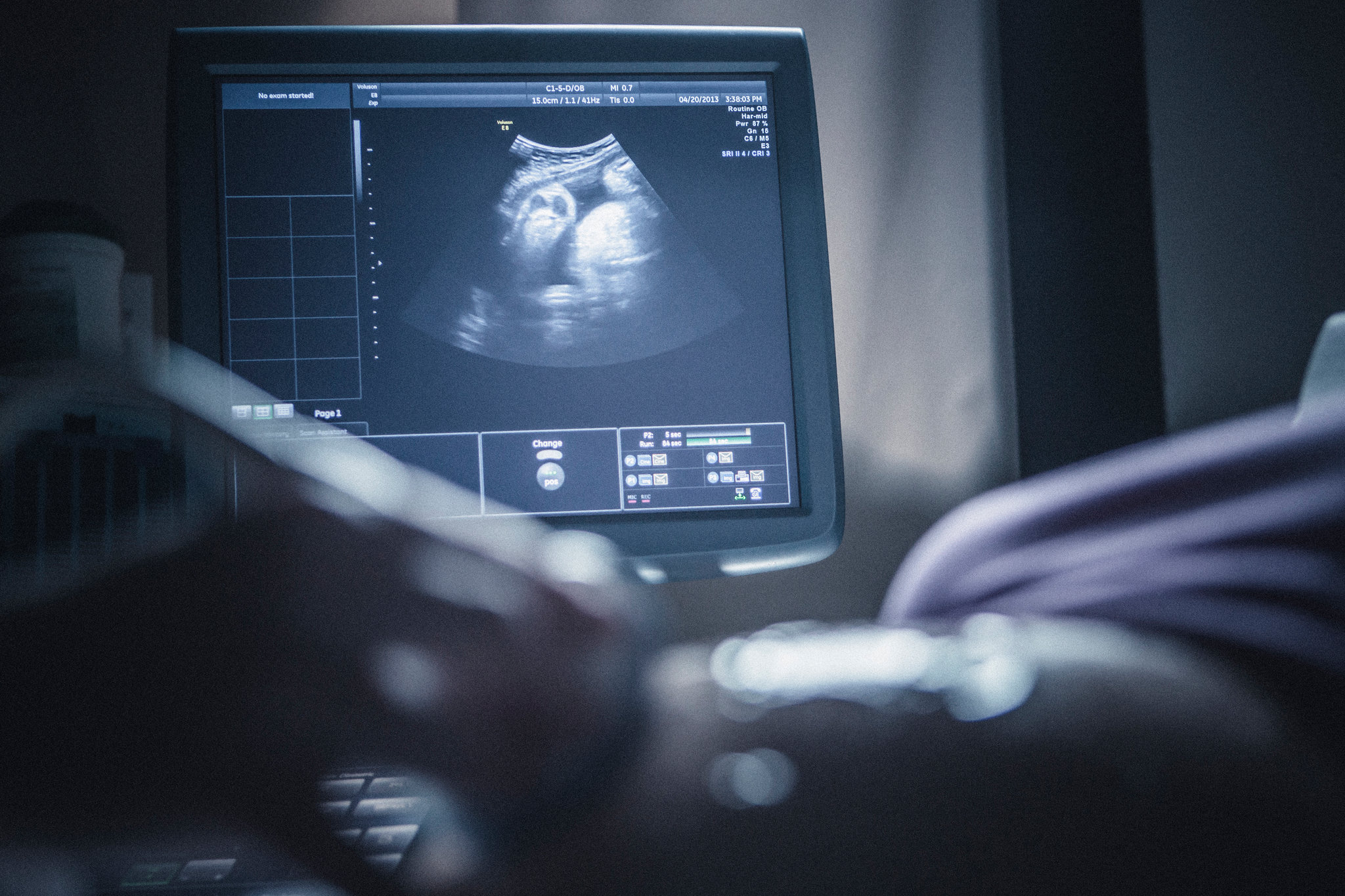
Ultrasound is a non-invasive imaging examination that uses sound waves to produce images of structures within the body. It can be used to examine different organs and tissues and may assist healthcare professionals in evaluating a patient's condition.
Professional Diagnostic ServiceWe provide a range of ultrasound examinations covering different areas of the body and different diagnostic needs.
Ultrasound examination used to assess the female reproductive organs and related pelvic structures.
Ultrasound examination used during pregnancy to support the assessment and monitoring of pregnancy.
Examination of pelvic structures to provide diagnostic information for appropriate clinical assessment.
Ultrasound examination of abdominal organs and structures to support clinical evaluation.
Ultrasound examination covering abdominal and pelvic structures through a transabdominal approach.
Examination of structures associated with the urinary system to support appropriate clinical assessment.
Ultrasound examination of breast tissue to provide additional diagnostic information where clinically appropriate.
Ultrasound examination of muscles, tendons, joints and other musculoskeletal structures.
Ultrasound examination performed through a transvaginal approach to assess relevant pelvic and reproductive structures.
Ultrasound examination performed through a transrectal approach for appropriate diagnostic assessment.
Ultrasound examination of the thyroid gland to support evaluation of thyroid structure and related findings.
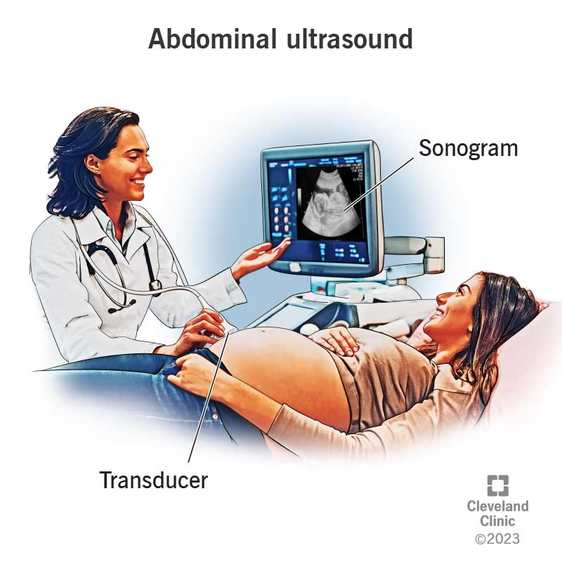
Ultrasound examinations can provide valuable diagnostic information and may be requested as part of a patient's medical evaluation. The appropriate examination depends on the patient's symptoms, clinical history and the request or referral from a qualified healthcare professional.
Preparation requirements may differ depending on the type of ultrasound examination. Patients should follow the instructions provided by the facility or their healthcare provider before attending their examination.
Providing laboratory and ultrasound diagnostic services with a commitment to professionalism, reliability and patient-focused care.
The Ultimate Scientific Prove
